 |
|
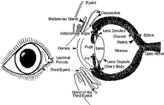
Eye definitions |
|
|
|
|
|
|
|
Aqueous Humour: Is a watery fluid that fills the front of the eye and maintains the pressure / firmness of the globe. The fluid also provides the nutritional support for the cornea and lens. It is produced inside the eye by the ciliary body and processes, and drains out through the iridocorneal angle.
Cornea: The cornea is the clear circular window on the front of the eye. It is made up of 3 basic layers. The centre layer is the stroma – a dehydrated fibrous layer like a dry sponge, with inner and outer water proofing layers (endothelium and epithelium). There should be no blood vessels in the cornea – erosions of the corneal surface are called ulcers.
Iridocorneal Angle. This is the drainage angle for the out flow of aqueous humour out of the eye, and is between the cornea and the base of the iris. Abnormalities of the drainage angle can predispose to increased pressure inside the eye (glaucoma).
Iris: The iris is the coloured part inside the eye, usually brown, but may be green, orange or blue. It is a muscular sheet which sits behind the cornea and in front of the lens, and acts like a camera’s diaphragm with a changing aperture. This helps controlling sharp focus and the amount of light reaching the retina.
Lacrimal Puncta. These are the 4 openings for the drainage of tears, in most pets they are positioned on the inside upper and lower eyelid margins.
Lens: The lens is a transparent, nearly spherical ball inside the eye, which sits behind the iris and is surrounded by an elastic capsule. It is like a plastic bag full of raw egg white, which can change shape to assist with fine focussing. Opacities or cloudiness in the lens are usually called cataracts.
Optic Disk: The optic disk is a spot on the retina where the tracks (fine nerves) from the photoreceptors all meet up to make the optic nerve.
Optic Nerve: The optic nerve is essentially the cable of sensory nerves from the eyes retina / photoreceptors to the brain.
Pupil: The pupil is the black hole in the centre of the iris which light passes through. The size of the pupil is controlled by the iris muscles. The pupil will be larger in low light and smaller in bright light.
Retina: The retina is essentially the light receptor for vision (like a solar cell or the film of a non-digital camera). When stimulated by light, the retina sends electrical impulses to the brain which are interpreted as vision.
Sclera: The sclera is the tough white fibrous coat of the eye.
Tapetum: This is a coloured reflective layer under the retina giving better night vision to those animals which possess them (not in man).
Uvea: The uvea is the middle vascular / blood vessel layer of the eye, and is divided into the iris (coloured part of eye), ciliary body and choroid. Uveitis is the inflammation of these parts of the eye.
Vitreous Humour: The vitreous is a clear jelly like liquid that fills the majority of the back portion of the eye (from the back of the lens to the retina).
Zonules: The zonules are the support structures of the lens, holding it in place. They are made up of 100s of fine string like fibres, attaching from the ciliary processes to the lens capsule.
|
|
What do animals see? |
|
|
|
|
|
|
|
Visual Acuity & Colour perception
One of the more common questions presented to us is: how well do animals see, can they see in colour? These are actually quite difficult questions to answer accurately; pets can’t read our colour or Snellen eye charts. However it is possible to make some educated assumptions about there vision by studying their behaviour, position of their eyes in the skull and anatomy of their eyes.
The Retina & Colour Vision
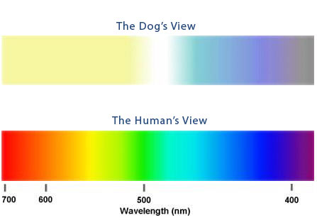
The retina is essentially the light receptor for vision (like a solar cell or the film of a non-digital camera). It contains two basic types of vision receptor cell (photoreceptors) called rods and cones. Basically the rods are best for low light conditions and motion detection, while the cones are for bright light conditions, small objects and colour appreciation. Humans have very high levels of cones compared to most animals (nearing 100% in the central retina compared to 20% cones in the dog). Both Humans and cats have three types of cone receptor, while dogs have two. Evidence suggests dogs have colour vision similar to humans with red-green colour-blindness as shown below.
|
|
|
|
Visual acuity |
|
|
|
|
|
|
|
Visual acuity is the ability to see two separate objects as separate, rather than blurring into one. The normal visual acuity in dogs is estimated at about 20-40% that of Humans. (For example a dog could identify an object at 20 metres that a human could distinguish at 90 metres).
The actual acuity is often measured in “cycles per degree” with the following being reported results for various species
Species Cycles per degree (visual acuity)
People 30
Horses 18
Dogs 12
Cats 6
Light Sensitivity
The dog and cat visual systems are adapted for better function under low light conditions compared with humans. They have rod rich retinas, large corneas, and a tapetum (which acts like a reflector under the retina giving increased chance of any light being picked up by the retinas). The tapetum gives animals their particular “eyes-shine” often seen at night.
Humans do not have a tapetum, so rather than a yellow / green eye-shine, the back of the human retina is usually seen as red (which is the cause of the “red eye” fault in photos taken with flash photography).
|
|
Fields of view (Cats/Dogs/Equine) |
|
|
|
|
|
|
|
The position of the eyes within the orbit and head determines the total field of view, the amount of peripheral vision, and the amount seen by both eyes simultaneously – binocular vision. It is this binocular vision which gives accurate distance judgement. In general prey animals have increased peripheral vision (better scanning the horizon for potential risks), and predatory animals increased binocular vision (better quarry localization).
|
|
|
|
Can my pet cope blind? |
|
|
|
|
|
|
|
The loss of vision is most unfortunate for any patient, but most pets adjust well to their blindness and can lead a happy and fulfilled life. Their hearing, whiskers and particularly sense of smell are markedly better than ours and are more important for their fulfilment and quality of life.
Blind horses can be more problematic, however in a safe paddock with good fencing and a friendly companion – many cope very well.
Cats tend to become slightly more sedate and wander less, but rarely do they become lost. Blind cats are frequently found jumping and climbing fences and trees. Getting another companion pet that the blind patient can follow and interact with often greatly increases their enjoyment of life. Blind pets rapidly learn to negotiate their territory with ease without bumping into objects; they tend to follow the same tracks around their property, often following walls inside.
There are a few simple aids that can make your blind pets lives significantly easier.
- Keep their environment as safe and stable as possible, avoiding changes as much as possible. If you need to move furniture or into a new house, you may find it takes a while (4-6 weeks) for your pet to familiarize its self with the new environment.
- Keep their bed, food/water bowl and litter tray (if appropriate) in the same place. These areas become a reference site should your pet become disorientated. Applying a little perfume on the legs of furniture can help with their location.
- Your pet can be exercised but should always be supervised especially in unfamiliar surroundings, teaching them to walk on a lead or harness is safest with the “extender leads” being very helpful. Using more voice commands, teaching them ”no”, “stop”, ”sit” and “wait”, quickly on command can be very helpful. Play toys that are noisy and odorous are often popular.
- You should try to educate any visitors, especially small children about your pet’s blindness so they can announce their presence and approach without startling them.
- Both cats and dogs will benefit from a tag on their collar saying “I am blind and possibly lost – please call my owner”.
- Take care around pools, ponds and rivers, as they can fall in, become disorientated and may drown (even previous “water dogs” that could swim well when visual).
- A blind pet’s eyes should not discharge nor become red or painful, do not delay in having them examined by a veterinarian if this occurs.
Please also check out these websites that you might find useful
www.blinddogs.com Good introductory site
www.valianttrust.org story of a blind horse that performs dressage with his owner
|
|
What happens if my pet needs an eye removed? |
|
|
|
|
|
|
|
It is sometimes necessary to remove a pet’s eye due to irreversible, blinding and painful conditions. The surgery is called an enucleation. This procedure quickly eliminates discomfort and the need for medications which may be expensive and may be harsh on the whole body. During the procedure the entire eyeball is removed, and the eyelids are permanently sewn shut.
Once healing has taken place and the hair has grown back, an enucleation leads to a relatively cosmetic and rapidly pain free outcome. Before enucleation, we will discuss all possible treatment options for your pet.
|
|
|
|
 |

)
