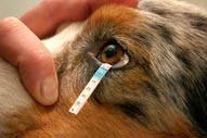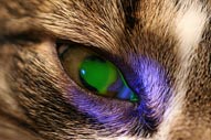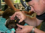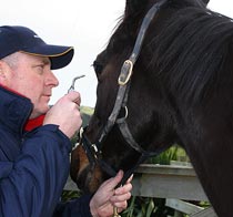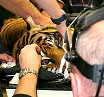
Fluorescein stain: This is yellow / green stain used for examining the surface of the eye – the stain attaches to scratches and ulcers on the corneal surface allowing evaluation of the size, severity and progression of the injury. It may also be used to assess the normal tear flow out of the eyes into the nose / mouth. |
|
Rose Bengal stain: This is another examining stain, which attaches to areas of more subtle damage to cornea and conjunctiva. |
|
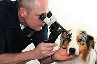
Biomicroscopy: Or slit lamp biomicroscopy is a valuable examining instrument for highly magnified assessment of the front part of the eye (cornea, iris and lens) and eyelids. |
|
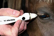
Tonometry: This is the measurement of the pressure inside the eye, and is measured with a “Tonometer”. A high pressure indicates glaucoma, a low pressure may indicate inflammation within the eye. |
|
Gonioscopy: This is a specialised examination of the internal drains inside the eye. The technique requires a special contact lens to be placed on the cornea. Gonioscopy is an aid in the diagnosis of, and predisposition to glaucoma. |
|
Ophthalmoscopy: This is the visualisation and assessment of the back of the eye (the retina / fundus) using an ophthalmoscope. Good communication between your veterinarian and the ophthalmologist is crucial for optimal patient care. Peter will telephone/ fax/ email a full report to your veterinarian within 24 hours after your pet has been examined and treated. |
|

